All our staff are BSHAA certified and covered by our comprehensive medical indemnity insurance.
Our technical staff includes fully qualified audiologists who are on hand to assess any issue which may arise during the initial ear examination.
The process begins with a client history where we discuss the procedure and alleviate any concerns. As well as an outer ear examination the technician will use a fibre optic otoscope to check the inside of both ear canals and the health of the tympanic membrane. If the canal and tympanic membrane are considered healthy then the technician will proceed to the next step in the process.
Ear Wax
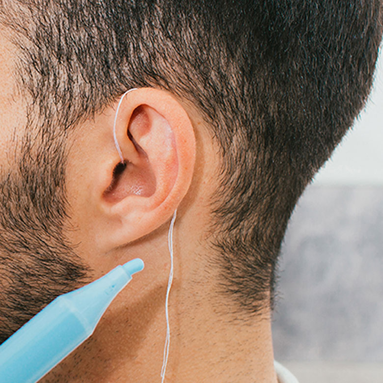
Ear wax can sometimes cause a problem when taking a scan of the ear so All our impression technicians are qualified to remove excess wax using micro-suction or water irrigation.
Cluistrom technicians are qualified and insured to remove ear wax which ensures a perfect scan or mould of the ear canal
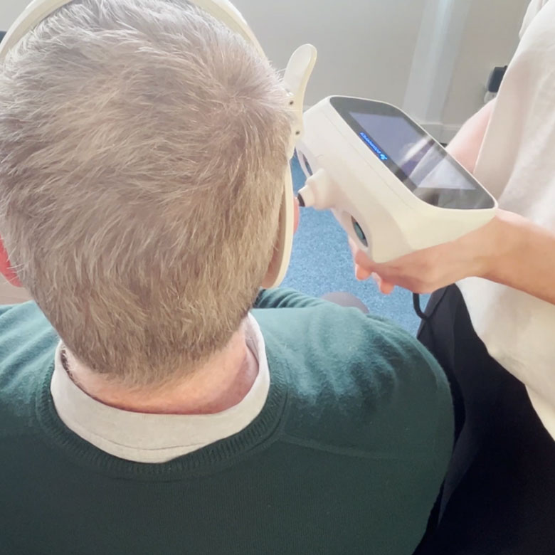
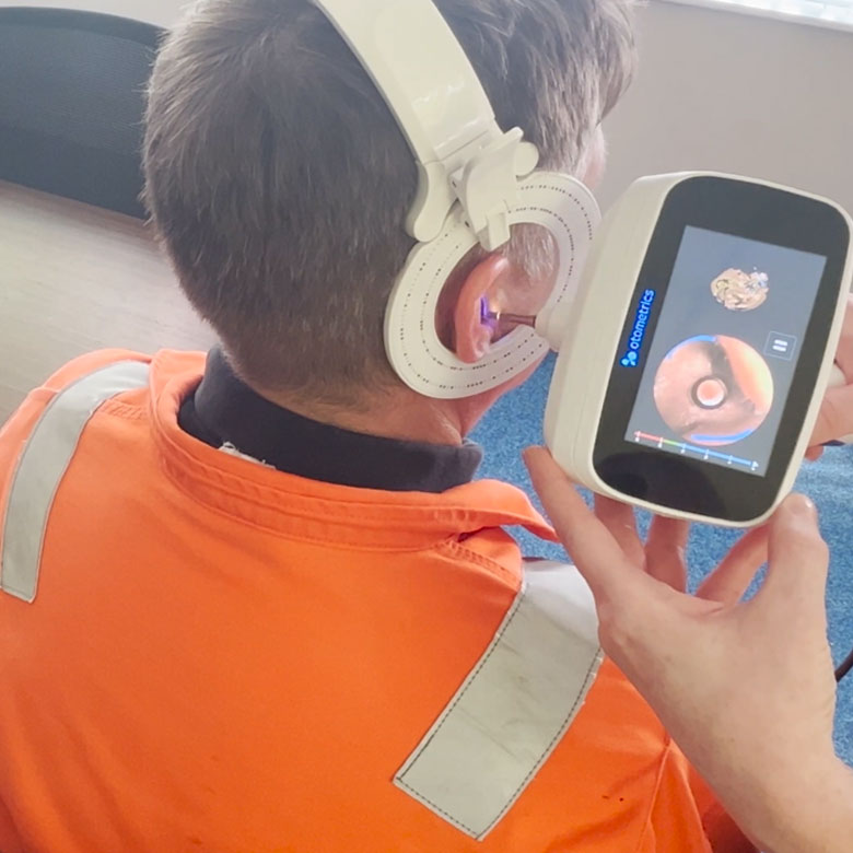
The technician will place the regulator headband on your head and use this to guide the laser marker around your ear canal and concha.
The laser has two operation modes-
Mode 1 uses a small ring laser that measures and maps the canal in a 360 degree process.
Mode 2 uses a separate laser with a horizontal beam that we use to map the outer cartilage of the ear and the concha (bowl). As the technician operates the laser scanner you will begin to see a new 3D image of your own ear and canal appear on the screen in front of you.
The resulting scans are labelled with a secure serial number and then sent via a secure network to the cloud server. Once stored and logged the scans can be downloaded for manufacture on one of the four new ProJet 3510MP Digital 3D Printers. This is often conducted overnight in a secure temperature and pressure controlled environment.
We make a great impression
This material is recognised as the most advanced available today.
Our unique process guarantees that the finished product is the truest likeness to the original impression taken and guarantees both comfort and acoustic performance over years of continued use.
Upon placing an order for custom moulded protection, one of our representatives will arrange a suitable date to visit your site to take the necessary ear impressions.
During the procedure our technician will check the health of the ear and advise or refer should there be a contra-indicator to performing a scan or cold impression
A sound survey can be carried out by fully qualified members of the Academy of Accoustics using Class 1 sound metres . This is an additional cost but ensures the correct filter strength is selected for the delivered product.
Your own existing sound survey can be used if there is one available .
After manufacture the products are supplied in their own secure carry case with full instructions and a cleaning tool. Lanyards are also available should they be required.
Make the leap from dissposable throw away earplugs and join some illustrious companies.
We provide earplugs and custom ear moulds to protect the hearing and prevent hearing loss for swimmers, hunters, musicians and many more.
Custom-Fit Earplugs To Isolate Yourself From Unwanted Sound.